 Macular degeneration (AMD) and cataracts play a major role in the United States health care system and global effort directed to the prevention of these conditions now is part of optometry initiatives. To benefit society both from a financial and a productivity perspective, optometrists focus on four areas in clinical practice: protective lenses, nutraceuticals, genetic testing and periodic examinations.
Introduction to disease prevention
In February 2014, approximately 24 optometrists gathered at the University of Houston for the first ever Ocular Surface Disease Wellness Conference. The subject of “wellness” and disease “prevention” were addressed in this historic two-day meeting. Prior to this meeting, most specialized gatherings by optometrists addressed disease diagnosis and treatment, rather than prevention. The concept of disease prevention is unique in the vision care arena. One possible exception is in the area of myopia where efforts to retard progression have been tried using bifocal eyeglasses, contact lenses (orthokeratology) and pharmacological agents (atropine). A follow-up meeting has been scheduled for December, 2014 in Dallas, Texas.
Two conditions that lend themselves to prevention discussions by optometrists in the United States include ocular surface disease (OSD) and ocular damage from high-energy visible light (HEV) as well as damage from ultraviolet light (UV). Macular degeneration (AMD) and cataracts play a major role in the United States health care system and any effort directed to the prevention of these conditions will benefit society both from a financial as well as a productivity perspective. Cataract surgery is the most commonly performed surgical procedure in the United States today. The average cost of cataract surgery today is $3,230 per eye, and it is rising because of the use of new technology (laser cataract surgery and multifocal IOLs). Estimates of the global cost of visual impairment due to age-related macular degeneration is $343 billion.
Specific to AMD prevention, U.S. optometrists now focus on four areas of preventative steps. Those areas include nutritional supplements, genetic testing, specialty lens coatings to block selective wavelengths of blue light, and periodic dilated fundus examinations with OCT studies. While there are several OCT models available, we have personally been pleased with our newer Cirrus™ HD-OCT instrument. Genetic risk assessment for age-related macular degeneration is becoming commonly employed in the United States for those patients with risk factors. Steve Arshinoff writes: “Previously we considered the phenotypic appearance of the eye, macular pigment levels and patient-related non-genetic factors to determine AMD risk.” Genotyping with commercially available genetic testing (Macula Risk™, RetnaGene™) now allows us to predict with 90 percent accuracy an individual’s 2-, 5- and 10-year risk for progression to advanced AMD. Following the reporting of the Age-Related Eye Disease Study 2 (AREDS2) (NEI) results, we now have definitive information on AMD prevention and progression using particular nutritional supplements, although further research is necessary.
Pathophysiology and economics
The number of people living with macular degeneration is similar to that of those who have been diagnosed with all types of invasive cancers. As many as 11 million people in the United States have some form of age-related macular degeneration. The number is expected to double to nearly 22 million by 2050. Most researchers believe that blue light exposure has a role in the pathogenesis of AMD. According to Margrain et al.: “Laboratory evidence has demonstrated that photochemical reactions in the oxygen-rich environment of the outer retina lead to the liberation of cytotoxic reactive oxygen species (ROS). These ROS cause oxidative stress, which is known to contribute to the development of AMD. The precise chromophore that may be involved in the pathogenesis of AMD is unclear but the age pigment lipofuscin is a likely candidate.”They continue: “Studies in human macular pigment density and the risk of AMD progression following cataract surgery lend further weight to the hypothesis that blue light exposure has a role in the pathogenesis of AMD but the epidemiological evidence is equivocal. Blue-violet light has a twofold effect on lipofuscin. It causes an increase in production and also activates its phototoxic components (free radicals), causing the death of RPE cells. On balance the evidence suggests but does not yet confirm that blue light is a risk factor in AMD.” Research by the Schepens Eye Institute (Harvard University) suggests that a low density of macular pigment may also represent a risk factor for AMD by permitting greater blue light damage.
Science

Existing artificial light sources are basically of two types: incandescent (includes halogen) and luminescent (fluorescent and LED). Incandescent lights are becoming difficult to find in the typical home repair stores in the United States as the newer LED light sources begin replacing them. These newer light sources are much more energy efficient, have a longer lifetime and the government has decreed that this exchange takes place. It is thought that by 2020, 90% of all light sources worldwide will be based on solid state lighting products and LEDs. These newer light sources give off a greater proportion of blue light than the older incandescent bulbs. We know that the sun is the standard light source. The blue light proportion of our daylight in the entire visible spectrum varies between 25% and 30%. We know that blue light is vital to a number of physiological processes and interfering with it may have adverse effects. A recent study by Gray and colleagues in the Journal of Cataract and Refractive Surgery found that patients with blue-light filtering IOLs performed significantly better under driving conditions with glare compared with similar patients who had clear IOLs. Dr. Henderson and her colleagues see no harm posed by blue filters, at least in visual parameters: they “feel that the potential protection against AMD is worth it.”
Clinical practice
Several groups around the world have studied the potential health risks of products using LEDs. Basically three high-risk populations have been identified: (1) children and aphakes who receive a higher blue light proportion on the retina, (2) those individuals suffering from ocular photosensitive pathologies or using photosensitive drugs (light-sensitive agents used in photodynamic therapy such as Verteporfin used to ablate blood vessels in the eye when treating wet macular degeneration), and (3) those individuals who are daily exposed to LEDs while using short viewing distances.
1. AMD AND PROTECTIVE LENSES
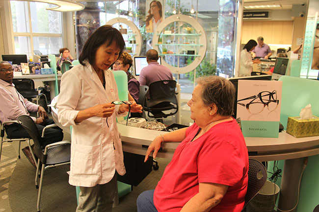
As a practical matter, optometrists and ophthalmologists in the United States have begun the process of utilizing electronic medical records (ObamaCare). During the early part of the patients’ examination they are asked several questions by the technician and those individuals who fall into one of the above three groups are then counseled on the particular risks they face and are prescribed spectacle lens treatments which will help protect them from the increased threats offered by increased blue light presence. Because optometrists are the guardian of good vision, it is important for us to counsel patients about modifiable risk factors. Two of those risk factors include smoking and cumulative light exposure, especially UV and HEV blue light.

We have found at the Clayton Eye Center in Morrow, Georgia that the best results are achieved when the doctor himself/herself initiates the conversation about blue light protection in the xamination room and then the dispensing optician reinforces the message. We specifically prescribe the new Crizal® Prevencia® lens treatment in order to selectively filter out only the dangerous wavelengths while allowing the good wavelengths to pass through. We know that blue wavelengths are the most potent portion of the visible electromagnetic spectrum for circadian regulation. Because the timing and quantity of light and darkness both affect sleep, evening use of amber lenses to block blue light might affect sleep quality. We have found over the past several months that our patients appreciate the fact that we are protecting their eyes with these discussions and we feel that to not educate our patients would be a great disservice. We reference data from the Beaver Dam and Blue Mountain studies, which implicate blue light as a factor for age-related macular degeneration, particularly following cataract surgery. Our clinic performs more than 3,000 cataract surgeries a year and each post-op visit emphasizes the potential risk of blue light. Our IOLs are blue blocking for added protection. Our practice recently became involved with an accelerated program emphasizing doctor-directed dispensing. Each of our nine optometrists now prescribes various lens products and coatings to each patient when indicated and outlines the specific products on a specially designed form and reviews these products with the patient. The patient is then escorted to the optical department from the clinical area by a technician or the doctor and the form is presented to the dispensing optician. Products such as AR, Transitions®, digitally surfaced progressive designs (such as the Essilor S Series ™) and Crizal® Prevencia® coatings have increased substantially as a result of this new process.
Several lens manufacturers have become involved with blue blocking technology; however, so far only Essilor has designed a coating that blocks specific wavelengths. VSP’s Unity BlueTech lens, Hoya’s Recharge ™ , PFO’s iBlucoat ™ and Signet Armorlite’s BlueTech (Indoor and Outdoor) all block HEV (high energy visible light) and offer improved contrast sensitivity. However, they also block the blue-violet range, which has been demonstrated to be “good” light and necessary for other functions, including increased contrast sensitivity and mood regulation.
2. AMD AND NUTRACEUTICALS
The Age-Related Eye Disease Study 2 (AREDS2) was a multi-center, randomized trial designed to assess the effects of oral supplementation of macular xanthophylls (lutein and zeaxanthin) and/or long-chain omega-3 fatty acids (docosahexaenoic acid) [DHA] and eicosapentaenoic acid [EPA]) on the progression to advanced age-related macular degeneration (AMD). While the results of the study left several questions unanswered, it also led the way to changes with respect to the prescribing of nutritional supplements for those patients with early macular degeneration and those at risk. Optometrists in the United States now routinely encourage their patients to take these nutraceutical supplements as a matter of procedure and this author suspects that this practice will become standard of care in only a matter of time. There are several commercial products on the market to choose from: Bausch and Lomb’s Preservision Eye Vitamin AREDS 2 Formula Soft Gels are probably the most commonly used. This particular product is beta-carotene free, which is a positive for current/former smokers. Another product that I have frequently used is Science Based Health’s Macula Protect Complete, which is also beta-carotene free. The study demonstrated that there was a 25% overall risk reduction of progression to exudative AMD. The role of macular pigment (MP) is also acknowledged and many optometrists now measure macular pigment and dose supplements accordingly. The U.S. diet is known to be low in lutein and zeaxanthin. The third carotenoid, Meso-Zeaxanthin, is a key carotenoid in the macula and even lower in the U.S. diet. We know that smokers are at high risk for AMD. In smokers and former smokers, betacarotene has been associated with an increased risk of lung cancer.

3. AMD AND GENETIC TESTING
Genetic testing has progressed in several areas of medicine over the past ten years. One area that has enjoyed the benefits of continuing research is AMD. We can now make a prognosis to within 90% accuracy of how a patient’s eye disease will progress. Several research projects have demonstrated that those patients subjected to testing have better outcomes than those without. At the 2013 American Society of Retina Specialists Annual Meeting, Dr. Peter Sonkin, a retina specialist from Tennessee Retina, presented the results from an analysis of the impact of genetic testing in their practice over a five-year period. The data revealed that patients who had Macula Risk testing and were subjected to a stratified surveillance schedule as well as a patient education program had better visual acuities on presentation compared to those patients without genetic testing. The November 2013 Issue of Ophthalmology highlighted an article titled “Prediction of Age-Related Macular Degeneration in the General Population – The Three Continent AMD Consortium”, which is a study evaluating AMD prognostics using three prospective population-based studies: the Rotterdam Study, the Beaver Dam Eye Study, and the Blue Mountain Eye Study. The non-genetic model which included age + sex + BMI + smoking + AMD status has a 78% predictive accuracy, while the genetic model, which included genetics with the above criteria, had an 82% predictive accuracy. Using all available information I have now come up with a formula for what the primary care optometrist should now do to prevent vision loss. This protocol is used by our doctors and many others and is a compendium of existing good practices.
4. THE CLAYTON EYE CENTER VISION LOSS MODEL IN AMD
- Diagnose AMD
- Perform genetic testing on each AMD patient
- Increase monitoring frequency including OCT testing
- Prescribe the appropriate nutraceuticals
- Prescribe selective blue-blocking spectacle lenses
- Counsel patients on diet, smoking, exercise and weight (BMI).
Optometrists in the U.S. have embraced genetic testing for AMD much in the same way other physicians have embraced genetic testing for cancer and several other diseases. There are over 2,000 tests available. Screening embryos for disease is becoming more frequent.
Conclusion
In summary, AMD is on the rise. As individuals continue to live longer, the optometrist is going to diagnose increasingly larger numbers of cases. Prevention is a must. We now have several tools that will allow us to aid in our preventative efforts. Government-mandated lighting changes will expose us to larger doses of potentially harmful HEV blue light. Computer usage continues to be on the rise and these tools as well as electronic tablets, smart phones and other games used at closer near point distances will also increase our exposure. By prescribing spectacle lenses that can aid in filtering out the noxious wave lengths, we may be able to prevent many individuals from acquiring this dreadful disease down the road. By adding genetic testing and nutraceutical supplements to our armamentarium, we may be doing the world a tremendous favor. Our job is vision preservation, and this is one way to accomplish that task. To not implement the above protocol but rather take a “wait and see” approach may be doing your patients more harm than good. The Optometric Oath promoted by the American Optometric Association includes the following charges:
- “I WILL advise my patients fully and honestly of all which may serve to restore, maintain or enhance their vision and general health.
- I WILL strive continuously to broaden my knowledge and skills so that my patients may benefit from all new and efficacious means to enhance the care of human vision.”
- The above approach fulfills my responsibility.
Article from the magazine "Point de vue"


