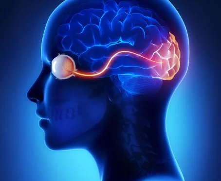
Vision a collaboration between eyes and brain
 Vision is neither present in the eyes nor in the brain, but results from the collaboration between the eyes and the rest of the brain. Vision is an omnipresent aspect of our existence, permeating all our activities.
It develops through neural plasticity and can be improved. Optometry is the discipline dedicated to the management of all aspects of the visual process.
INTRODUCTION
The fact that vision seems so effortless belies the complexity of the visual process. The term vision refers to the complex of eye and brain.1 It is this complex which guides a broad spectrum of human abilities.
We walk down a street, step up and down curbs, maneuver around objects and other pedestrians and adjust our pace, while visually monitoring our position. Moments later we get into our car, drive at highway speed through traffic and judge where we are relative to other vehicles while anticipating the flow of traffic. We arrive at baseball practice where we pick up a bat, walk to the plate, miss a curve ball, foul-off a fast ball and then hit a single, making numerous conscious and subconscious judgements with varying degrees of success. After practice, we stop by the mall, scan the crowd for our friend, and go to the bookstore to find the book we might want to purchase. Over a no-foam latte, we read the opening chapter, seeing if it captures our attention.
Our vision plays an essential role in each of these activities through the collaboration of eyes and brain.
VISUAL NEUROSCIENCE
The retina is a thin sheet of brain tissue in the eyes. It is the place where the brain first encounters light.2 Signals travel back-and-forth between the eyes and the rest of the brain. Visual processing involves several major sub-cortical centers plus a mosaic of dozens of distinct areas in the cerebral cortex.10 It is currently acknowledged that vision is the result of parallel, distributed processing in multiple areas and through multiple pathways.11 For example, information gathered by the retina about color is processed in a different area of the brain than information about movement.4
As another example, egocentric direction describes the perceived location of an object compared to our body. This is derived from a combination of oculocentric direction (where our eyes are aimed), position of the eyes in the head, and the head’s position relative to the body. The brain uses a reference point midway between the two eyes, known as the egocenter to compute egocentric direction.12 Enabling the brain to engage a whole body experience, binocular vision is an intricate organization of biologic and psychologic components.7
The majority of nerve cells from the retina project to the visual cortex. However, at least ten percent of the nerve cells take a different pathway8 stimulating areas of the brain stem dedicated to functions that seem remote to vision, when vision is narrowly defined.9 The existence of extensive sensory motor pathways supports a broader conceptualization of vision, integrating functions such as balance and visual-auditory localization.13
“As surely as the old system (for explaining vision) considered that the problem of knowledge and understanding could be separated from the problem of seeing, so the present one will find it increasingly difficult to draw a dividing line between the two.”3
CLINICAL OPTOMETRIC SCIENCE
Many aspects of the optometric examination probe the eye-brain collaboration. Consider the complexity of the evaluation of visual fields. A patient is instructed to simultaneously maintain steady central fixation, attend to central and peripheral stimuli, discriminate threshold stimuli, and demonstrate awareness with an appropriate motor response.
Similarly, the clinical assessment of color vision requires more than the discrimination of colors.
Every color test has multiple perceptual components. For example, color vision plates require recognition of form and the emergence of figure from background. Color cap tests (Farnsworth) are predicated on good sequencing abilities and subtle discriminatory skills.
The process of binocular vision is a reflection of complex interactions within the eye-brain continuum. For neural binocular summation to occur, inputs from both eyes to the brain must be synchronized in both space and time.5 Alignment of the eyes is maintained via ongoing collaboration of eyes and brain. Binocular dysfunctions such as suppression and anomalous correspondence demonstrate cortical adaptations in the eye-brain function to minimize visual confusion and maintain some level of visual performance.6
It is evident that, beyond eye health, optometrists evaluate a wide variety of visual abilities.
These include visual-spatial orientation skills, visual analysis skills (including auditory-visual integration, visual discrimination, visual figure-ground perception, visual closure, visual memory, and visualization), visual motor integration skills, and visual-verbal integration skills.14
CONCLUSION
Information from neuroimaging and insights from cognitive neuroscience demand a significant reformulation of the understanding of vision. Vision occurs neither in the eyes nor in the brain, but emerges from the collaboration of the eyes and the rest of the brain. Vision is a pervasive aspect of our existence which permeates all of our activities. Vision develops and, due to neural plasticity, can be enhanced. Optometry is the discipline dedicated to the care of all aspects of the visual process.
This publication was formulated by the American Optometric Association’s Binocular Vision
Working Group. The following individuals are acknowledged for their contributions:
Gary J. Williams, O.D., Chair
Gregory Kitchener, O.D.
Leonard J. Press, O.D.
Glen T. Steele, O.D.
Approved by: American Optometric Association, April 2004
REFERENCES
1. Marmor MF, Ravin JG The eye of the artist. St. Louis: Mosby 1997
2. Sterling P, How retinal circuits optimize the transfer of visual information, In; LM Chalupa, JS Werner, eds. The visual neurosciences. Cambridge, MA: The MIT Press, 2004
3. Zeki S, A Vision of the Brain. Oxford: Blackwell Scientific Publications 1993, p. 144
4. Van Essen DC, Gallant JL, Neural mechanisms of form and motion processing in the primate visual system. Neuron 1994; 13: 1-10
5. Steinman SB, Steinman BA, Garzia RP. Foundations of binocular vision: A clinical perspective. NY: The McGraw-Hill Companies 2000
6. Caloroso EE, Rouse MW. Clinical management of strabismus. Stoneham, MA: Butterworth-Heinemann 1993
7. Harwerth RS, Schor CM. Binocular vision. In: Adler’s physiology of the eye. 10th ed. PL Kaufman, A Alm, eds. St. Louis: Mosby, 2003.
8. Bear MF, Connors BW, Paradiso MA. Neuroscience: Exploring the brain. Baltimore, MD: Williams & Wilkins, 1996
9. Bartley SH. The human organism as a person. Philadelphia, PA: Chilton, 1967
10. Van Essen DC, Deyoe EA. Concurrent processing in the primate visual cortex. In: MS Gazzaniga, ed. The cognitive neurosciences. Cambridge, MA: The MIT Press. 1995
11. Cassagrande VA, Xu X. Parallel visual pathways: A comparative perspective. In: LM Chalupa and JS Werner, eds. The visual neurosciences. Vol. 1 Cambridge, MA: The MIT Press. 2004
12. von Noorden GK, Campos EC. Binocular vision and ocular motility: Theory and management of strabismus. 6th ed. St. Louis: Mosby 2002
13. Soderquist DR. Sensory Processes. Thousand Oaks, CA: Sage Publications, 2002
14. Optometric Clinical Practice Guideline. Care of the Patient with Learning Related Vision Problems. St. Louis: American Optometric Association 2000
15. Zenger B, Sagi D. Plasticity of low-level visual networks. In: M Fahle and T Poggio. Perceptual Learning. Cambridge, MA: The MIT Press. 2002
Article from the American Academy of Optometry

